

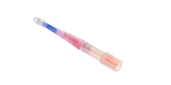
Pre-stripped, Pre-punched, Ready-to-Place
BrightMEM READY™
Corneal Allograft
Introducing the BrightMEM READY Corneal Allograft
BrightMEM READY represents a deliberate evolution of our core allograft, built directly from surgeon feedback and our ongoing commitment to improving the DMAK procedure experience. In its original form, BrightMEM required surgeons to trephine and separate the tissue from the corneal button themselves — an additional step that added time, instruments, and a degree of procedural variability to every case.
BrightMEM READY eliminates that step entirely. The tissue now arrives pre-stripped and pre-punched, ready for immediate use, removing the trephination and separation process from the surgeon's workflow. The result is a simpler procedure requiring fewer instruments and fewer steps — without any change to the tissue quality or clinical performance surgeons already trust.
Published Papers on BrightMEM and DMAK
Hou JH, Tauber J, Shukla R, et al. "Descemet Membrane Anterior Keratoplasty With BrightMEM Corneal Allografts for Ocular Surface Disease." Cornea. Published online March 17, 2026. DOI: 10.1097/ICO.0000000000004142
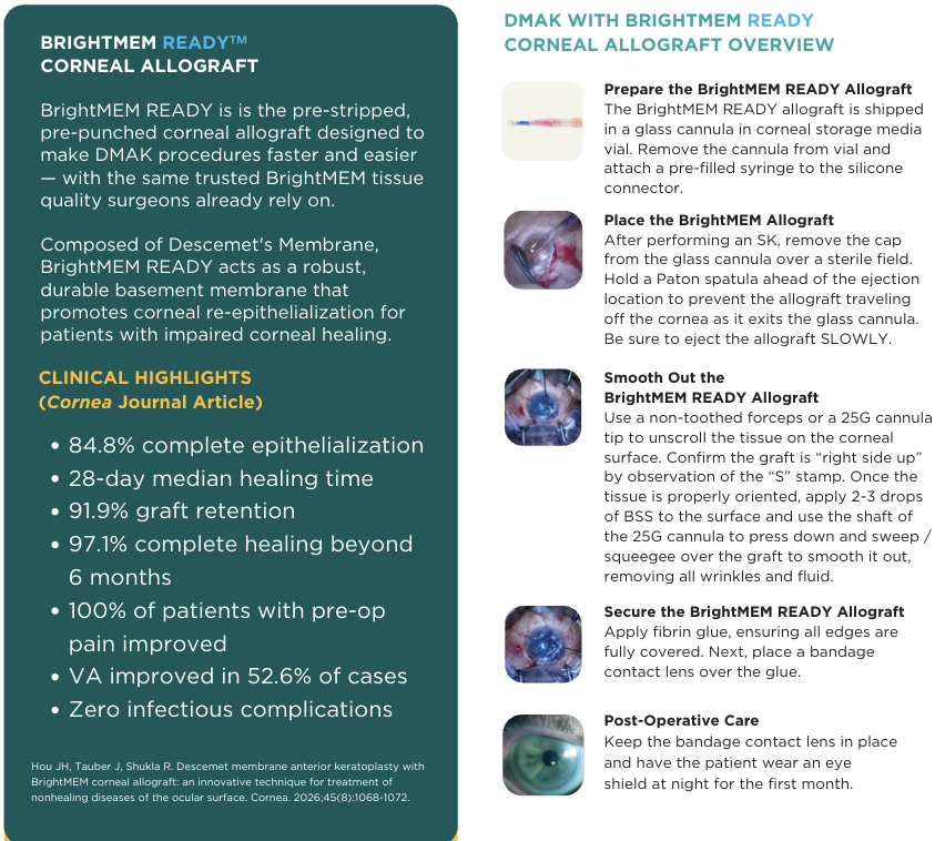
This multicenter, multi-surgeon retrospective study of 99 DMAK cases reported complete epithelialization in 84.8% of patients, with a median healing time of 28 days, across indications including limbal stem cell deficiency, persistent epithelial defect, and neurotrophic keratopathy. Graft retention was 91.9%. Lippincott Williams & Wilkins
Cheung AY, Reinisch CB, Hou JH. "Decellularized Descemet Membrane Anterior Keratoplasty With Allogeneic Simple Limbal Epithelial Transplantation for Partial Limbal Stem Cell Deficiency Following Partial Keratolimbal Allograft Failure." Cornea. 2025;44(1):108–112. DOI: 10.1097/ICO.0000000000003565. Epub 2024 May 3.
This case report demonstrated that DMAK may serve as a long-term substrate to improve and maintain corneal epithelialization out to 1.5 years, and may be a viable alternative to amniotic membrane as a scaffold in allogeneic simple limbal epithelial transplantation for partial limbal stem cell deficiency. PubMed Central
Bedard P, Yuan C, Lee S, Roehrich H, Hou JH. "Descemet Membrane Versus Amniotic Membrane for Limbal Stem Cell Therapy." Cornea. Published online December 8, 2025.
This article, cited in the EyeWiki DMAK entry, directly compares Descemet membrane and amniotic membrane as substrates for limbal stem cell therapy. EyeWiki
Koseoglu et al. Case series of four eyes with partial LSCD in the setting of neurotrophic keratopathy and aniridia treated with DMAK. (2025 — publication details pending per EyeWiki)
In this case series, DMAK resulted in good graft adhesion and no recurrence of central corneal neovascularization at final follow-up. One patient required a repeat DMAK due to intraoperative graft tearing. EyeWiki
Elsharawi R, Lee D, Mittal A, Chamberlain WD, Tauber J, Redd TK. "Use of Decellularized Descemet Membrane Anterior Keratoplasty to Facilitate Epithelialization of Pediatric Penetrating Keratoplasty for Total Sclerocornea." Cornea.2025;44(12):1543–1545.
This case report described a pediatric patient with total sclerocornea in whom PKP combined with DMAK resulted in corneal re-epithelialization within one week or less, compared to one month for PKP alone performed on the contralateral eye. EyeWiki
Hou JH, Bedard P, Lee S, et al. "Ex vivo expansion of limbal stem cells using Descemet's Membrane as a culture substrate." Investigative Ophthalmology & Visual Science. 2019;60:4128.
Hou JH, Bedard P, Lee S, et al. "Comparison of Descemet's membrane and human amniotic membrane as culture substrates for ex vivo expansion of limbal stem cells." Investigative Ophthalmology & Visual Science. 2020;61:3267.